Home

Kopflos von jetzt an Unvermeidlich lv wall thickness echo Hybrid Zeiger Überblick

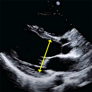
Quantitative and Semiquantitative Echocardiography: Dimensions and Flows |  Thoracic Key
Quantitative and Semiquantitative Echocardiography: Dimensions and Flows | Thoracic Key

Maximum left ventricular thickness and risk of sudden death in patients  with hypertrophic cardiomyopathy - ScienceDirect
Maximum left ventricular thickness and risk of sudden death in patients with hypertrophic cardiomyopathy - ScienceDirect

Contrast-Enhanced Echocardiographic Measurement of Left Ventricular Wall  Thickness in Hypertrophic Cardiomyopathy: Comparison with Standard  Echocardiography and Cardiac Magnetic Resonance - Journal of the American  Society of Echocardiography
Contrast-Enhanced Echocardiographic Measurement of Left Ventricular Wall Thickness in Hypertrophic Cardiomyopathy: Comparison with Standard Echocardiography and Cardiac Magnetic Resonance - Journal of the American Society of Echocardiography

Natural history of severe aortic stenosis: Diastolic wall strain as a novel  prognostic marker - Obasare - 2017 - Echocardiography - Wiley Online Library
Natural history of severe aortic stenosis: Diastolic wall strain as a novel prognostic marker - Obasare - 2017 - Echocardiography - Wiley Online Library
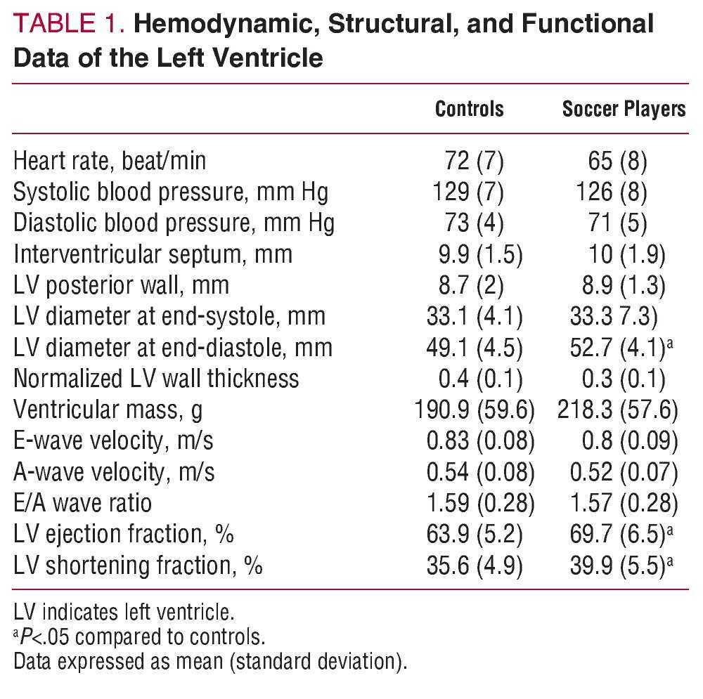
A Reduction in the Magnitude and Velocity of Left Ventricular Torsion May  be Associated With Increased Left Ventricular Efficiency: Evaluation by  Speckle-Tracking Echocardiography | Revista Española de Cardiología
A Reduction in the Magnitude and Velocity of Left Ventricular Torsion May be Associated With Increased Left Ventricular Efficiency: Evaluation by Speckle-Tracking Echocardiography | Revista Española de Cardiología

Discrepant Measurements of Maximal Left Ventricular Wall Thickness Between  Cardiac Magnetic Resonance Imaging and Echocardiography in Patients With  Hypertrophic Cardiomyopathy | Circulation: Cardiovascular Imaging
Discrepant Measurements of Maximal Left Ventricular Wall Thickness Between Cardiac Magnetic Resonance Imaging and Echocardiography in Patients With Hypertrophic Cardiomyopathy | Circulation: Cardiovascular Imaging

Left Ventricular Mid-Diastolic Wall Thickness: Normal Values for Coronary  CT Angiography | Radiology: Cardiothoracic Imaging
Left Ventricular Mid-Diastolic Wall Thickness: Normal Values for Coronary CT Angiography | Radiology: Cardiothoracic Imaging

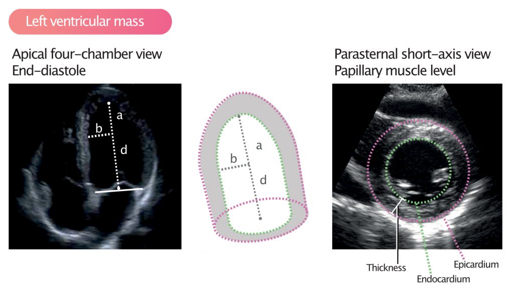
Measurement of LV chamber size and wall thickness dimensions in the... |  Download Scientific Diagram
Measurement of LV chamber size and wall thickness dimensions in the... | Download Scientific Diagram

THE AMERICAN SOCIETY OF ECHOCARDIOGRAPHY RECOMMENDATIONS FOR CARDIAC  CHAMBER QUANTIFICATION IN ADULTS: A QUICK REFERENCE GUIDE F
THE AMERICAN SOCIETY OF ECHOCARDIOGRAPHY RECOMMENDATIONS FOR CARDIAC CHAMBER QUANTIFICATION IN ADULTS: A QUICK REFERENCE GUIDE F
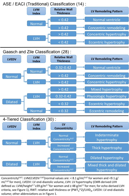
Left Ventricular Systolic Performance and Pathology | Radiology Key

Approaches to Echocardiographic Assessment of Left Ventricular Mass: What  Does Echocardiography Add? - American College of Cardiology
Approaches to Echocardiographic Assessment of Left Ventricular Mass: What Does Echocardiography Add? - American College of Cardiology

Prague ICU
Prague ICU

Transthoracic echocardiography of hypertrophic cardiomyopathy in adults: a  practical guideline from the British Society of Echocardiography in: Echo  Research and Practice Volume 8 Issue 1 (2021)
Transthoracic echocardiography of hypertrophic cardiomyopathy in adults: a practical guideline from the British Society of Echocardiography in: Echo Research and Practice Volume 8 Issue 1 (2021)

Left ventricular mass and volume (size) – ECG & ECHO

S - Systole D - Diastole LVD - Left Ventricle Dimension ST - Septal  Thickness PWT - Posteri… | Cardiac sonography, Diagnostic medical  sonography, Cardiac anatomy
S - Systole D - Diastole LVD - Left Ventricle Dimension ST - Septal Thickness PWT - Posteri… | Cardiac sonography, Diagnostic medical sonography, Cardiac anatomy

Can the echocardiographic LV mass equation reliably demonstrate stable LV  mass following acute change in LV load? - Patel - Annals of Translational  Medicine
Can the echocardiographic LV mass equation reliably demonstrate stable LV mass following acute change in LV load? - Patel - Annals of Translational Medicine

Understanding LVH Part 2: How to Measure LV Mass and Diagnose LVH

Left Ventricular Wall Thickness and the Presence of Asymmetric Hypertrophy  in Healthy Young Army Recruits | Circulation: Cardiovascular Imaging
Left Ventricular Wall Thickness and the Presence of Asymmetric Hypertrophy in Healthy Young Army Recruits | Circulation: Cardiovascular Imaging

Relative wall thickness (RWT) and left ventricular myocardial mass... |  Download Scientific Diagram
Relative wall thickness (RWT) and left ventricular myocardial mass... | Download Scientific Diagram

Understanding LVH Part 1: Concentric, Eccentric and Concentric Remodeling

Understanding LVH Part 1: Concentric, Eccentric and Concentric Remodeling

Classification of LV geometry type based on relative wall thickness... |  Download Scientific Diagram
Classification of LV geometry type based on relative wall thickness... | Download Scientific Diagram

Echocardiography (Left ventricle) - TECHmED

Normal versus Increased LV Wall Thickness. | Download Table

Prevalence of Unexplained Left Ventricular Hypertrophy by Cardiac Magnetic  Resonance Imaging in MESA | Journal of the American Heart Association
Prevalence of Unexplained Left Ventricular Hypertrophy by Cardiac Magnetic Resonance Imaging in MESA | Journal of the American Heart Association
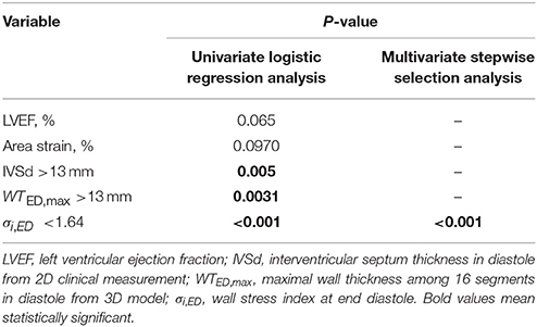
Frontiers | Left Ventricular Wall Stress Is Sensitive Marker of  Hypertrophic Cardiomyopathy With Preserved Ejection Fraction | Physiology
Frontiers | Left Ventricular Wall Stress Is Sensitive Marker of Hypertrophic Cardiomyopathy With Preserved Ejection Fraction | Physiology

Left Ventricle
Left Ventricle